Burns Imaging
Burns Imaging: Friend or Foe?
Burns are considered one of the most life-threatening of injuries. More so because of the likelihood and extent of burn injury in the very young, and the very old.
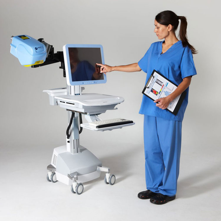
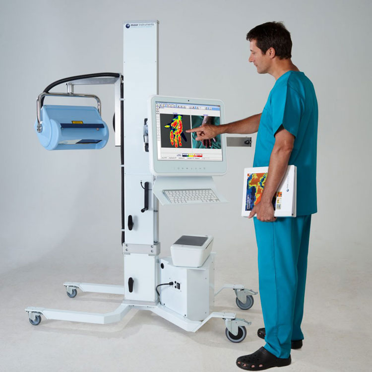
Burns Imaging is achieved by using a laser doppler imager. There are two models available in Australia which are both TGA/ARTG approved for use in burns imaging. These are the moorLDLS-BI and the moorLDI2-BI. Each model has its own advantages (and limitations) however both produce highly accurate scan images.
In a recent publication by Hoeksema et al the moorLDLS-BI was clinically evaluated in a multi-centre paediatric study, with excellent results. The group concluded that “The high accuracy of the new line-scan imager was comparable to that of the traditional LDI. Its size and mobility enabled easier ward and outpatient use. The higher scan speed was particularly beneficial for scans in paediatric patients*.”
Laser Doppler Burns Imaging FAQs:
Q: How does laser doppler imaging work:
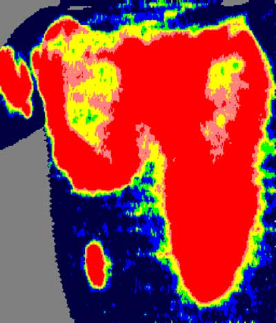
A. Laser light penetrates the skin and is scattered by tissue and by moving blood cells in capillaries, arterioles and venules. The moving blood cells in the tissue cause Doppler frequency shifts, which are then processed to produce a colour coded map of flow across the skin.
A digital camera records a colour clinical photograph at the same time, corresponding closely with the blood flow image in size and aspect. The measurement is non-contact and can quantify differences in flow over an area of tissue.
Q: After a burn injury has occurred, when should scanning be performed?
A. Scanning is recommended between 48 hours and 5 days post-burn.

MoorLDI2-BI image of wounds predicts most will heal within 14 days
Q: How long does a scan take to complete?
A. It depends upon the scan size and resolution, but typically between 40 seconds and 1 ½ minutes for a moorLDI2-BI scan and typically 4-8 seconds for a moorLDLS-BI scan. The duration of a scan session will depend upon the number of scans required to assess all of the burn(s).
Q: How deeply does the laser light penetrate?
A. As the skin and microvasculature are not homogenous this can be a difficult question to answer. Approximately 2 mm is a good estimation. However, this has not been found to limit burn assessment accuracy in areas where skin is thicker (e.g. the back).
Q: Are the moorLDI2-BI and moorLDLS-BI DICOM compatible?
A. Yes, DICOM compatible versions are available. Please contact us for additional information.
Q: What is the largest area I can scan?
A. The largest area in a single scan is approximately 50cm x 50cm for a moorLDI2-BI scan and 15cm x 20cm for a moorLDLS-BI scan, approximately the size of an adult hand. The moorLDLS-BI has a repeat scan mode which enables multiple scans to be taken across larger burns.
Q: Is laser doppler imaging contraindicated in pregnancy?
A. No.
Q: Why would I choose to use Laser Doppler Imaging?
- Early and accurate burn diagnosis from 48 hours post burn – avoiding unnecessary delays
- Non-contact imaging – pain free assessment with improved infection control
- Instant results – enables prompt assessment and treatment planning within the burns unit
- Clinically proven software with touch screen interface – easy to operate by burns team staff following training
- Unique colour-coded palette – easy to interpret, proven accuracy in numerous published studies and clinical trials
- CE marked, 510k registered specifically for clinical burn diagnosis and recommended by National Institute for Health and Clinical Excellence (NICE) – independent evidence of suitability for purpose
Q: So, Friend or Foe?
A: Undoubtably Friend. Laser Doppler imaging is an especially useful tool that offers you the opportunity to improve assessment accuracy to more than 96% – increasing your confidence in treatment decisions.
Relevant Equipment:
moorLDLS-BI: Laser Doppler Line Scanning
moorLDI2-BI: Single Point Laser Doppler


Combined with Clinical Judgment, Laser Doppler imaging can improve assessment accuracy to more than 96%.
Links to source webpages
Links to any references, papers
Henk Hoeksema, Rose D. Baker, Andrew J.A. Holland, Travis Perry, Steven L.A. Jeffery, Jozef Verbelen, Stan Monstrey. A new, fast LDI for assessment of burns: A multi-centre clinical evaluation. Burns, In Press, Corrected Proof, Available online 1 July 2014
An audit of the use of LDI in the assessment of burn depth, by Pape et al. (2001), reported 97% accuracy with LDI compared to 60-80% accuracy for established clinical methods. This study also found that use of LDI enabled unnecessary surgery to be avoided, resulting in a reduction of both costs and workload.
Pape, S.A. Baker, R. D. Wilson, D. Hoeksema, H. Jeng, J.C. Spence, R.J. Monstrey, S (2012) Burn wound healing time assessed by laser Doppler imaging (LDI). Part 1: Derivation of a dedicated colour code for image interpretation Burns, Burns, 38, p187-94.

