Imaging and Microscopy
Whole Slide Imaging
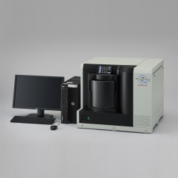
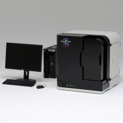
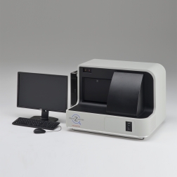
The NanoZoomer series is a family of whole slide scanners that convert glass slides into high-resolution digital data by high-speed scanning. The NanoZoomer comes with a variety of functions such as image acquisition of fluorescence samples and multilayer acquisition. Scanned data can be viewed on a PC monitor by using the dedicated viewer software, and patented navigation map technique* delivers slide viewing environment just as if operating a microscope. Advantages:
- Copying and Sharing: Digitized slides can be copied and shared. This feature of whole slide imaging can be used in a variety of applications. For example, a large group of people can observe and discuss a single sample.
- Slide Storage: Digital data does not deteriorate, and it is more secure from damages and losses than glass slides. You can view digital data in its original quality anytime and anywhere.
- Databases: Large numbers of whole slide imaging can be stored into a database and incorporated into a laboratory information system. You can share data and construct slide libraries with distant facilities and research institutes.
- Networks: Using the Internet or a local area network, you can observe and evaluate slides from a distant location.
Applications for Whole Slide Imaging include:
Routine & Computer Aided Diagnoses in the Pathology Laboratory. The ability to quickly produce and share high quality virtual slides paves the way for routine use of image analysis software to support the diagnostic decisions made by Pathologists. Advanced digital slide scanning systems automatically scan batches of slides and identify slides by their barcodes which frees Pathologists from the more mundane aspects of reporting and should also help to reduce errors. A fully digital medium enables an integrated approach to patient data collection, distribution, reporting and the seamless integration of different data types – images, text, database, statistics, quantitative analysis – into flexible information management systems.
Research. The use of Tissue Microarray Analysis (TMA) is now becoming standard practice in the research pathology laboratory. Digital slide scanning systems which can produce a virtual slide of a TMA slide allied with the use of TMA analysis software significantly increase the throughput of these slides, enabling data to be extracted and analysed more rapidly and efficiently.
Education & Training. Virtual microscopy allows viewing of virtual slides by large numbers of students or trainees over a computer network, thus avoiding the necessity of attending a teaching or training session. Traditionally, the students and trainees would have viewed images generated by a digital camera mounted on a microscope. Using web server software, it is now possible for each person to view a section of the slide selected by them at the magnification they choose. Not only are there significant cost savings to be made but the quality of the learning experience is enhanced.
Telepathology and Telehealth. The physical bulk of large slide collections and the area a pathologist may be responsible for is increasing and subsequently creating massive storage and accessibility problems. The creation of virtual slides which can be stored on a server, allowing access from anywhere in the world, will enhance the efficiency of a pathologist or department and reduce the risk of damaged or lost slides. Applications where this ability to access slides quickly and easily include cases for second opinions, MDT meeting preparation, intraoperative frozen sections, multisite clinical trials, education and training.

Key Products for Whole Slide Imaging:
Links to any references, papers


